 |
| Peter Riezebos Inverted Dogmatic Paradigm (1/3) |
~
Major
histocompatibility complex
(MHC) is the gene cluster that has immensely polymorphic character. MHC gene
cluster encodes MHC surface proteins which involve in antigen presentation
directly. Antigen-presentation is
serving a protein fragment to other relevant cells in order to create an
immunological response. This protein fragment presentation is highly important
for adaptive immunity and also it involves in triggering apoptosis of cells
that secrete misfolded protein fragment. Basically, MHC molecules are
glycoproteins that serve antigens to T cells. In human genome, MHC –gene
clusters are located on chromosome 6 and encodes α/β subunit proteins of MHC
molecules (Figure 1).
- Types and Structure") |
| Figure 1. Major Histocompatibility Complex (MHC)- Types and Structure |
There are mainly three groups of MHC molecules which is numerated as
MHC I, MHC II and MHC III1. In all nucleated cells, MHC I which, contains
3 α and β-microglobulin protein, is located on the cell surface and interacts
with cytotoxic T cells, CTLs (CD+8). Antigens that degraded in cytosol bind to
MHC I molecules without overhangs and resulted in apoptosis via CTLs. As
distinct from MHC I, intravesicular and extracellular antigens’ peptides bind
to MHC II molecules groove and it activates CD+4 T cells (Th) which stimulate
other immunological cells or mechanisms. So, MHC II molecules are present only
in professional antigen-presenting cells (APCs) such as dendritic cells, B
cells and macrophages. Peptide binding on MHC II groove is different from MHC I
in terms of being overhangs in both ends of peptide that binds to cleft2. Hence, antigens that bonded to MHC II molecules are
recognized by T Cell Receptors (TCRs) of CD+4 T cells. An additional
interaction between B7 protein & CD28 receptor is also needed for proper
activation (Figure 2).
 |
| Figure 2. Structure of MHC-peptide-TCR complexes. |
These sharp characteristic differences between MHC I & II
molecules dictate fate of response (Figure 3) by virtue of either endogenous or exogenous
degraded antigens3. Exogenous
antigens mainly bind to MHC II molecules, though. Sometimes MHC I molecules can
capture exogenous antigens which is described as unlawful (unexpected) usage of
class I molecules. Such phenomena involving exogenous antigen-MHC I interaction
are described as cross-presentation (Figure 4).
Cross-presentation is mainly occurred by dendritic cells (DCs) via their
endocytic/ phagocytic adaptions. During cross-presentation, two pathways are
named cytosolic and vacuolar pathway may
be chose depending on antigen processing4.
 |
| Figure 3. Antigen Presentation of MHC I & MHC II |
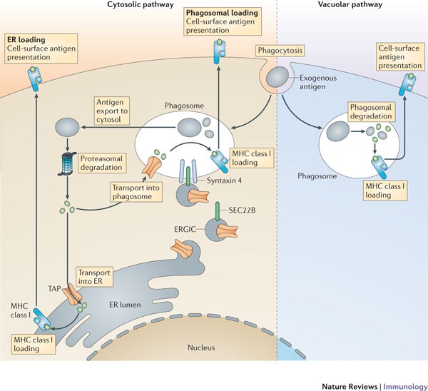 |
| Figure 4. Cross-presentation by dendritic cells. |
References
1. Rock, K. L., Reits, E., & Neefjes, J. (2016). Present Yourself! By MHC Class I and MHC Class II Molecules. Trends in immunology, 37(11), 724–737. https://doi.org/10.1016/j.it.2016.08.010
2. Neefjes, J., Jongsma, M. L., Paul, P., & Bakke, O. (2011). Towards a systems understanding of MHC class I and MHC class II antigen presentation. Nature reviews. Immunology, 11(12), 823–836. https://doi.org/10.1038/nri3084
3. Wieczorek, M., Abualrous, E. T., Sticht, J., Álvaro-Benito, M., Stolzenberg, S., Noé, F., & Freund, C. (2017). Major Histocompatibility Complex (MHC) Class I and MHC Class II Proteins: Conformational Plasticity in Antigen Presentation. Frontiers in immunology, 8, 292. https://doi.org/10.3389/fimmu.2017.00292
4. Joffre, O. P., Segura, E., Savina, A., & Amigorena, S. (2012). Cross-presentation by dendritic cells. Nature reviews. Immunology, 12(8), 557–569. https://doi.org/10.1038/nri3254
MUSTAFA ÖZTÜRK, revised from Immunology Course Assignments

Comments
Post a Comment